Photonis, an Exosens brand, in collaboration with the University of Leicester and European Space Agency (ESA), leveraging from its 40 years of experience in manufacturing of Microchannel Plates, has designed the Micro Pore Optics plate with square holes, to be used in X-Ray imaging and analysis applications. Micro Pore Optics (MPO) are routinely produced in our Photonis France manufacturing facility and are installed on a number of international space missions.
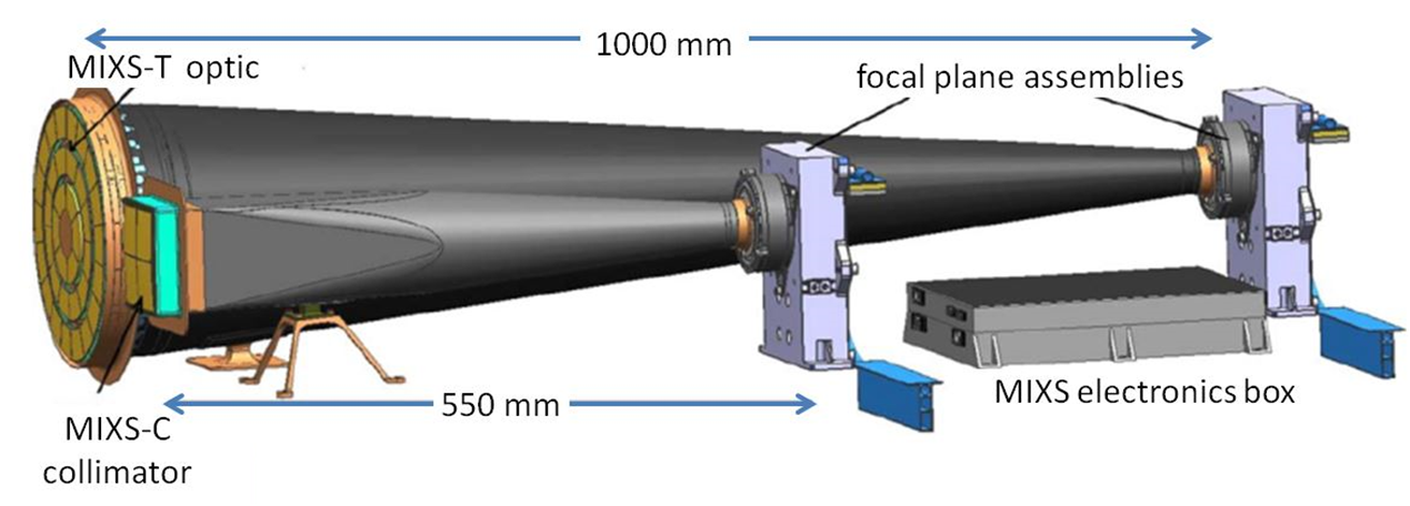
One of the examples, and the very first realization is for the MIXS-T and MIXS-C instruments for the currently flying BepiColombo mission to Mercury (under a joint ESA contract with University of Leicester).
The optics of the MIXS Mercury Imaging X-Ray Spectrometer entail:
a) a Wolter 1 type radial focusing “lobster eye” telescope: 2 radial optics assembled in series, each constructed of 3 concentric rings of spherically slumped radially packed MPOs, forming a mosaic of 72 MPOs with a focal length of 1m.
b) a collimating optic: a 2x2 matrix of spherically slumped square packed MPOs with a focal length of 55 cm.
For more information about BepiColombo mission and MIXS-T and MIXS-C instruments, visit websites below:
https://www.cosmos.esa.int/web/bepicolombo/mixs
https://le.ac.uk/bepicolombo/mixs
Principle of Operation
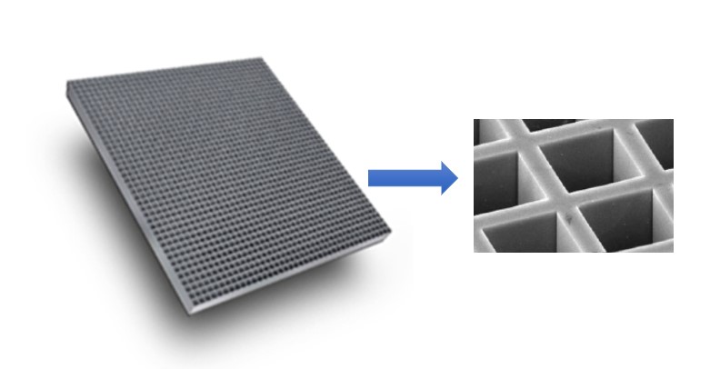
The Micro Pore Optics has millions of square channels, made by glass and often covered with a high reflective material such as Iridium, which act as mirrors and reflect the X-ray photons at very small grazing angles.
The Micro Pore Optics has millions of square channels, made by glass and often covered with a high reflective material such as Iridium, which act as mirrors and reflect the X-ray photons at very small grazing angles.
Its perfectly square and flat channels are optimized to allow X-Ray and UV photons to be focused, concentrated, or collimated due to the total external reflection. There are multiple possible configurations described below in which the MPO can be used for different purposes and offering multiple benefits for X-ray imaging and analysis.
Point-to-Point Focusing
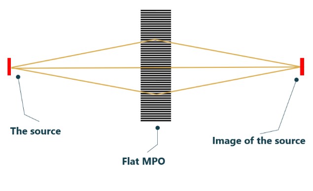
When a flat MPO is used as an X-ray optic, the incident photons from the X-ray source are focused after the MPO in a distance equivalent to the MPO-source distance, and configuration is called 1:1 or point-to-point focusing.
This configuration can be used to focus the divergent beam into another point, or to collimate the beam to a small size having much brighter spot compared to a pinhole collimator. MPO’s compact size allows to have a high degree of flexibility when placing the optics and use effectively the space in the set-up.
An example of use of focusing flat MPO is its successful integration by NASA SETI into prototype instruments MapX-I and MapX-II for planetary explorations, used for in-Situ 2D Mapping X-ray Fluorescence for the Detection of Biosignatures and Habitable Planetary environments.
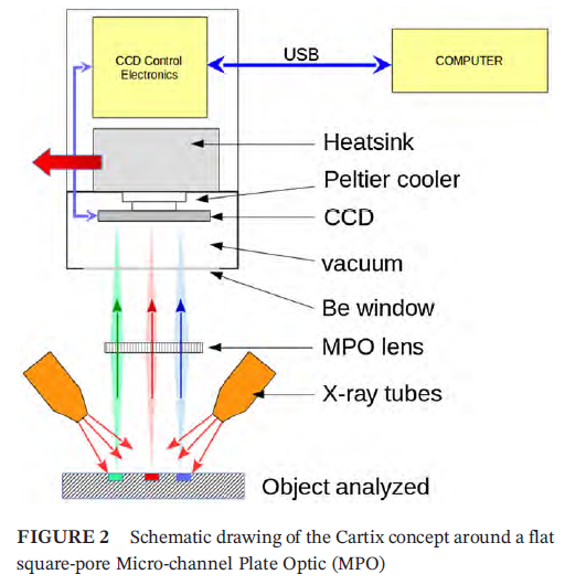
The same principal using a prototype instrument called Cartix was applied in non-destructive terrestrial applications e.g. to study Cultural Heritage such as paint composition of a French Impressionist painting and paint on Inca Pachacamac Idol Wood Sculptures in Lima, Peru.
Shown in a drawing, the Cartix is an arm-deployed Full-field X-ray fluorescence imaging (FF-XRF) spectrometer placed directly on a surface to be analyzed. A large area of a sample surface is irradiated by X-ray sources from both sides and the emitted XRF from the sample then collimated by a flat square pore MPO and guided to the CCD detector. This allows simultaneous acquisition of a 2D image of the fluoresced X-rays and rapid analysis in a 1:1 focusing configuration.
Credit Figure2: Philippe Walter, Philippe Sarrazin, Marc Gailhanou, Dominique Hérouard, Antoine Verney, David Blake
https://ntrs.nasa.gov/citations/20200001291
https://analyticalsciencejournals.onlinelibrary.wiley.com/doi/10.1002/xrs.2841
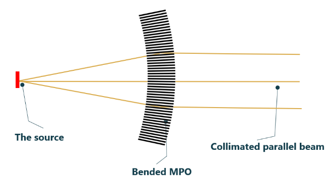
Slumped MPO Configuration: Parallel Beam
In many X-ray applications, for example in XRD for powder diffraction, thin film analysis, parallel beam is required with a very low divergence. Latter can be achieved when slumped MPO is used with an X-ray source placed in the focal point of the optics. MPO are available with a focal length from 50 mm, with high collection angles and compatible with large variety of X-ray sources.
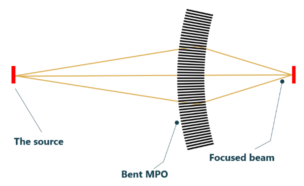
Slumped MPO Configuration: Focusing the Source
Slumped MPO can be also used to focus the X-rays, either parallel or divergent beams, to a much brighter spot. Whether it is for imaging or fluorescence applications, this allows obtaining very small size spots of few tens of microns with brightness much higher compared to pinhole collimators for example. Compared to other mostly very bulky optics, the MPO is the best alternative to solve the problems of space constraints allowing more flexible configurations for analysis of samples.